Blog Digital Health
Critical Care Suite 3.0: X-ray AI That Finds Feeding Tubes
On this page· 7 sections
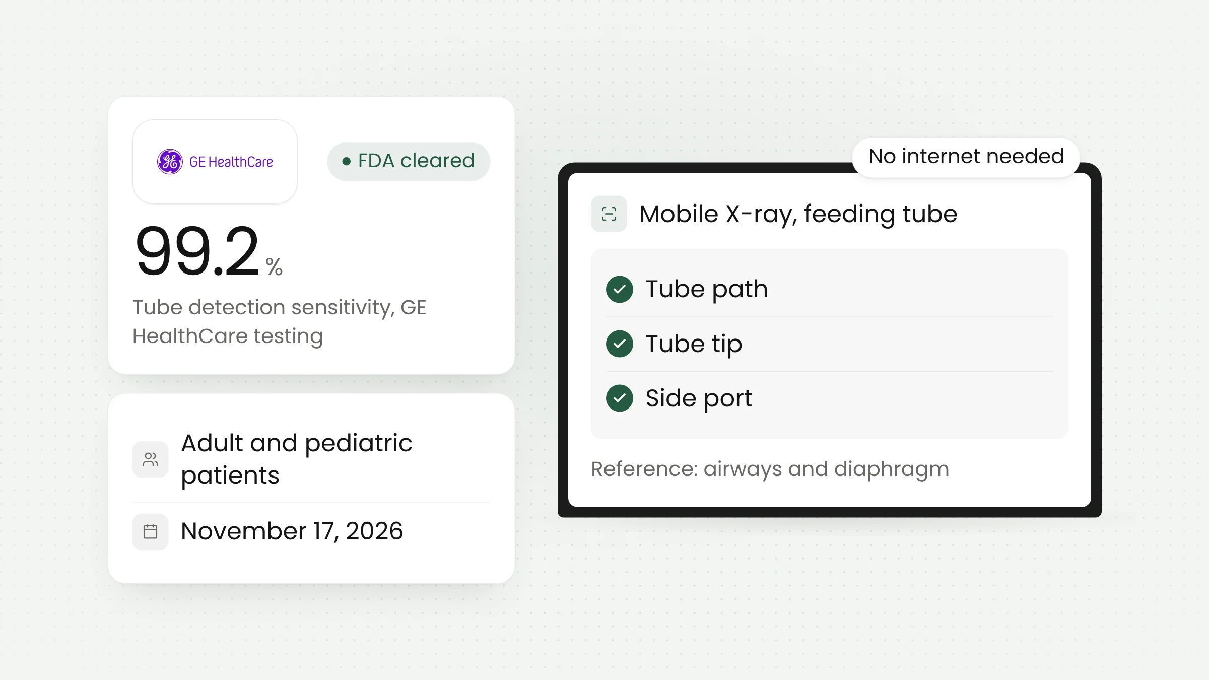
Critical Care Suite 3.0, GE HealthCare’s AI software for mobile X-ray systems, has received 510(k) clearance from the U.S. Food and Drug Administration. The company announced it on November 17, 2026. The new version detects feeding tubes on X-ray images of adult and pediatric patients, on the device itself and without an internet connection.
FDA clearance for feeding tube detection
According to the GE HealthCare press release, Critical Care Suite 3.0 is designed to automatically detect feeding (enteric) tubes on X-ray images and to support assessment of tube placement. The Chicago-based company describes it as the first on-device AI for that purpose in X-ray imaging. The submission is listed as 510(k) K253502.
A 510(k) clearance is the FDA pathway for medical devices judged substantially equivalent to a product already legally on the market. It applies to the United States.
Two things separate version 3.0 from its predecessors. The feeding tube algorithm is a new capability in the suite, and it is the first time GE HealthCare has included pediatric patients in that feature. Trade publication 24x7 reported the same points on the day of the announcement.
Larry Boyd, Vice President and General Manager of Global X-ray at GE HealthCare, set out the reasoning in the release: “Critical care teams need to make timely clinical decisions in complex, rapidly changing environments”.
Why feeding tubes are checked by X-ray
A feeding tube is passed through the nose into the stomach or small intestine to deliver nutrition and medication to patients who cannot eat. GE HealthCare cites research by Nikandros and colleagues in the journal Critical Care (2006) estimating that 80% of intensive care unit patients receive nutrition this way.
A tube in the wrong place can cause serious harm. A case commentary on PSNet, the patient safety site run by the U.S. government agency AHRQ, describes a 13-year-old boy whose feeding tube migrated from the small intestine to the stomach and then into the esophagus. He developed aspiration pneumonia and had to return to the ICU.
The authors, Norma Metheny and Kathleen Meert, recommend radiographic confirmation before a blindly inserted tube is used for the first time, followed by bedside checks of tube position every four hours. When those checks raise doubt, they call a radiograph the best test for establishing where the tube sits.
For critically ill patients, that image is usually taken with a mobile X-ray unit wheeled to the bedside. Citing the IMV 2024 Diagnostic X-ray Market Outlook Report, GE HealthCare says 75% of mobile X-ray system usage is in emergency departments and 51% in intensive care units.
How Critical Care Suite 3.0 works
The algorithms in Critical Care Suite 3.0 are embedded in the mobile X-ray device. After an image is acquired, the software marks three things: the path of the tube, its tip and its side port, the opening near the tip where feed comes out. It also highlights the airways and the diaphragm as anatomical reference points for judging where the tube lies.
The whole process runs without internet or cloud connectivity. Patient images do not have to be sent to an outside server for analysis, and the result appears at the bedside.
GE HealthCare stresses that the software is intended to work alongside care teams. The AI marks and highlights, while the clinical decision, such as withdrawing or repositioning a tube, stays with clinicians.
Performance figures reported by GE HealthCare
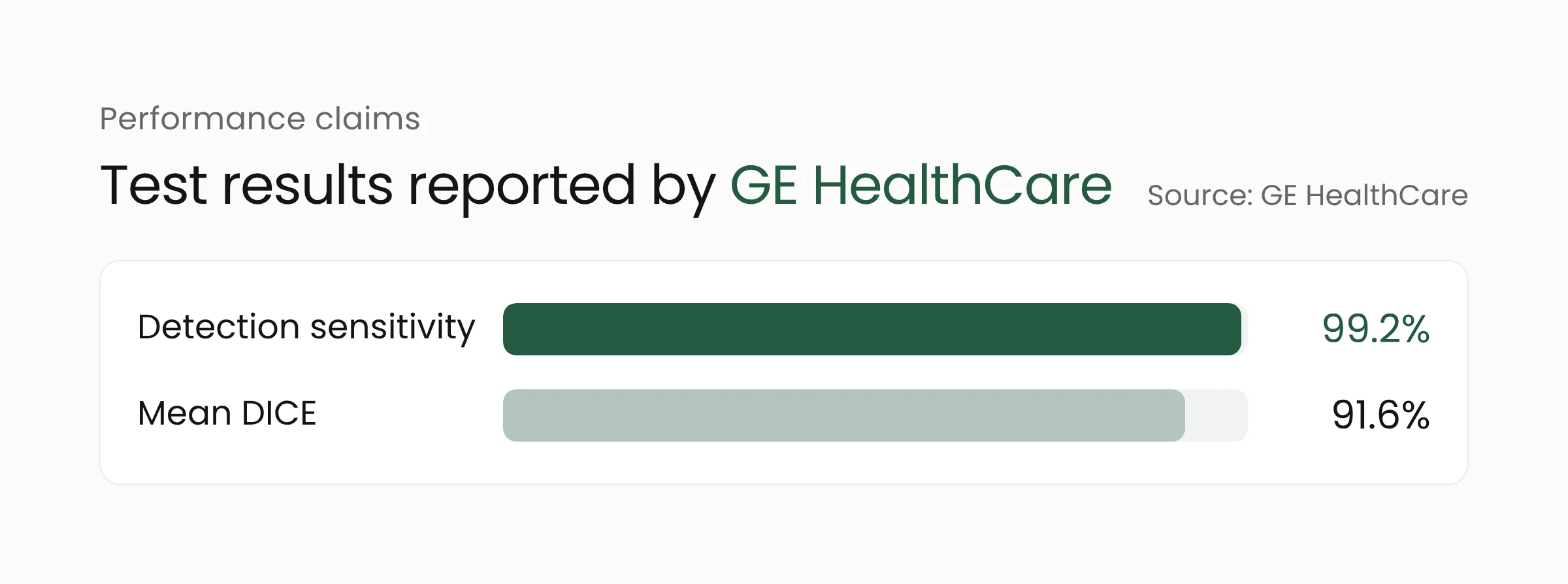
In testing reported by the company, Critical Care Suite 3.0 reached 99.2% sensitivity for tube detection. Sensitivity shows how many of the tubes actually present in the images were recognized.
The second figure is a 91.6% mean DICE score for localization. DICE measures the overlap between the area marked by the AI and the area marked by expert radiologists.
Both numbers come from GE HealthCare’s own testing and have not been verified by an independent party. The release also does not state how many images were tested, how results split between adults and children, or the specificity, which would show how rarely the AI marks a tube that is not there.
Three generations in five years
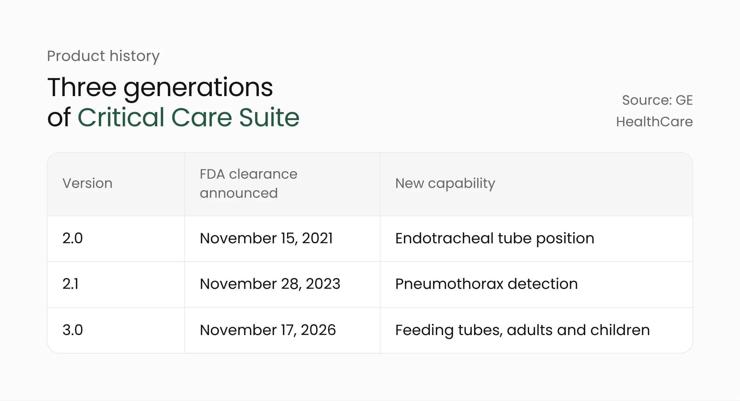
Critical Care Suite has been updated several times. On November 15, 2021, GE HealthCare announced FDA clearance for version 2.0 with an algorithm that measures the position of endotracheal tubes, the breathing tubes used for ventilated patients. That algorithm had been distributed since November 2020 under the FDA’s COVID-19 imaging guidance and was offered on the AMX 240 and AMX Navigate mobile X-ray systems.
On November 28, 2023, the company announced version 2.1 with an algorithm that detects pneumothorax, a condition in which air is trapped in the space around the lung.
Version 3.0 adds feeding tubes to that list. The package also includes Quality Care Suite, a set of algorithms that GE HealthCare says is aimed at reducing image quality errors.
Open questions after the announcement
The version 3.0 release names no supported X-ray models, no price and no country-by-country schedule. GE HealthCare only notes that product availability may vary by country. Availability in Indonesia and the rest of Southeast Asia has not been announced.
Industry outlet Zenopa writes that the feature is delivered as a software upgrade to the existing mobile X-ray platform, so hospitals would not need to buy new equipment. That detail does not appear in the official release.
The only user testimony comes from Adam Walbrun, clinical manager of imaging services at Emplify Health in La Crosse, Wisconsin, who said his team gets feeding tube assessment information at the bedside. GE HealthCare itself adds that the statement reflects one customer’s opinion and that results elsewhere may differ.
What this means for hospitals
Hospitals in Indonesia cannot buy this feature today. The useful part is the pattern it represents: AI built into the device, assigned one narrow task and independent of the network. We covered another FDA-cleared medical AI with a single specific task in our piece on AEYE-DS retinal screening.
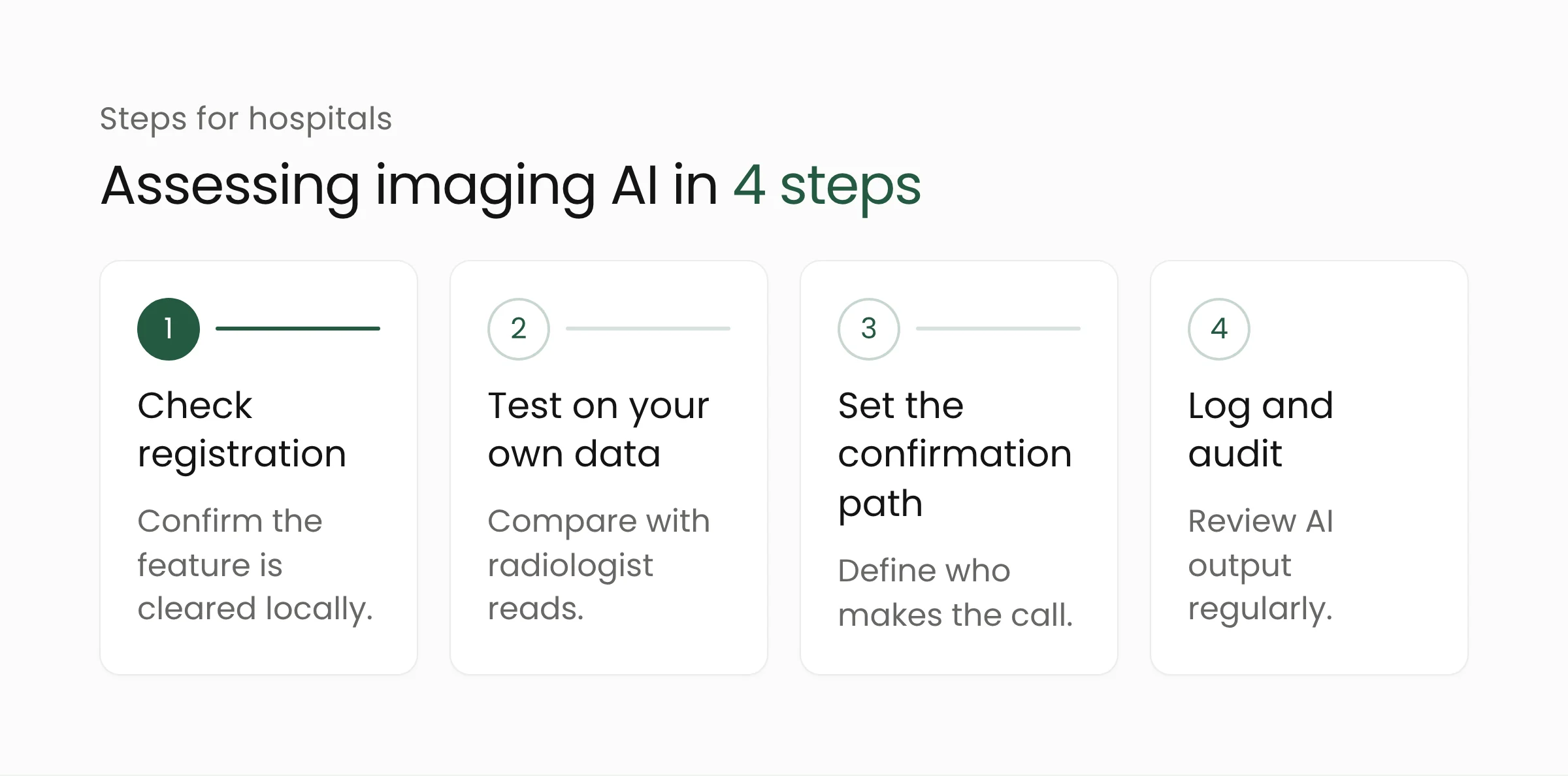
Management and radiology teams can apply the following four steps when weighing a similar offer from any vendor.
- Check registration. FDA clearance does not carry over to other countries. In Indonesia, medical devices, including their software features, need a marketing authorization from the Ministry of Health. Ask the vendor to show that the AI feature on offer is covered by it.
- Test on your own data. The 99.2% figure comes from the vendor’s test data. Before routine use, compare the AI output with radiologist reads on a set of your own patients’ images, including children and images crowded with lines and cables.
- Set the confirmation path. Write into procedure who is authorized to declare a tube position safe and when a radiologist must still read the image. An AI marker helps nurses and on-call doctors see faster, but it does not replace a radiologist’s expertise.
- Log and audit. Record the cases where the AI and clinicians disagree and review them regularly. That record is the basis for deciding whether the feature is worth keeping.
On-device architecture has particular value for regional hospitals. An unstable internet link does not stop the analysis, and patient images stay inside the building. The trade-off is that model updates depend on the vendor’s software release schedule, so the maintenance contract should spell that out.
AI output also needs to reach the medical record and the radiology system so it can be traced. For version 2.0, GE HealthCare stated that measurements and an annotated image were sent to PACS, the radiology image archive. Integration work of this kind is part of the healthcare solutions Merakit builds with care providers.
Frequently asked questions
Is Critical Care Suite 3.0 available in Indonesia?
Not announced. The clearance is an FDA clearance for the U.S. market, and GE HealthCare states that product availability may vary by country. Interested hospitals should ask the company’s official representative about local registration status.
Does this AI replace radiologists?
No. GE HealthCare says the software is intended to work alongside care teams. The AI marks the path, tip and side port of the tube on the image, while the final assessment and any action are decided by clinicians under hospital procedure.
Why run the AI on the X-ray unit and not in the cloud?
According to GE HealthCare, algorithms embedded in the device work without internet or cloud connectivity, so the result is available at the patient’s bedside. For hospitals, the approach also means patient images do not have to leave for an outside server to be analyzed.
Closing
Critical Care Suite 3.0 adds one narrow task to the list of jobs AI can assist with in intensive care. Its performance claims still need to be shown outside vendor testing, and its availability in Indonesia is unclear. If your hospital is weighing AI for imaging or other clinical workflows, Merakit’s artificial intelligence services team can help design the pilot and the integration.
Sources
- GE HealthCare’s Critical Care Suite 3.0 brings first on-device AI for feeding tube detection to X-ray imaging in adult and pediatric care, GE HealthCare, November 17, 2026
- GE HealthCare Gets FDA Clearance for Mobile X-Ray AI That Detects Feeding Tubes, 24x7, November 17, 2026
- FDA Clears GE HealthCare Mobile X-Ray AI for Feeding Tube Assessment, Respiratory Therapy, November 17, 2026
- GE HealthCare Lands FDA Clearance for On-Device Feeding Tube AI, Zenopa, November 19, 2026
- GE Healthcare X-ray AI receives FDA clearance to assess endotracheal tube placement, GE HealthCare, November 15, 2021
- GE HealthCare expands on-device triage capabilities with FDA clearance of algorithm for pneumothorax detection, GE HealthCare, November 28, 2023
- Case commentary on a migrated feeding tube, AHRQ PSNet, September 1, 2008